6+ Diagram Of Thoracic Vertebrae
These vertebrae form the foundation of the thoracic regions sturdy spinal column that supports the neck above the rib cage soft tissues flexible joints blood vessels and nerves. Web pedicle length decreases from T1 to T4 and then increases again as you move distal in the thoracic spine.
![]()
Illustration Of Thoracic Vertebrae Showing Vertebral Body Pedicles Download Scientific Diagram
Between the vertebral bones are disks that provide cushioning for your vertebrae and flexibility for.

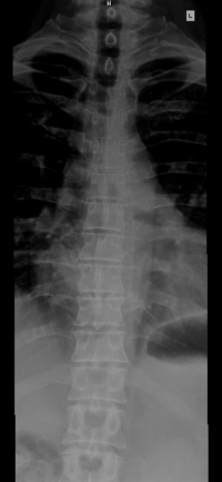
. Web Click To View Large Image. It consists of the 12 pairs of ribs with their costal cartilages and the sternum Figure 651 65. The thoracic spinal nerve 6 passes through underneath T6.
The thoracic spinal nerve 7 passes through underneath T7. The thoracic spine lies between the superior cervical spine and the inferior lumbar spine. Most of the thoracic vertebrae share various anatomical characteristics.
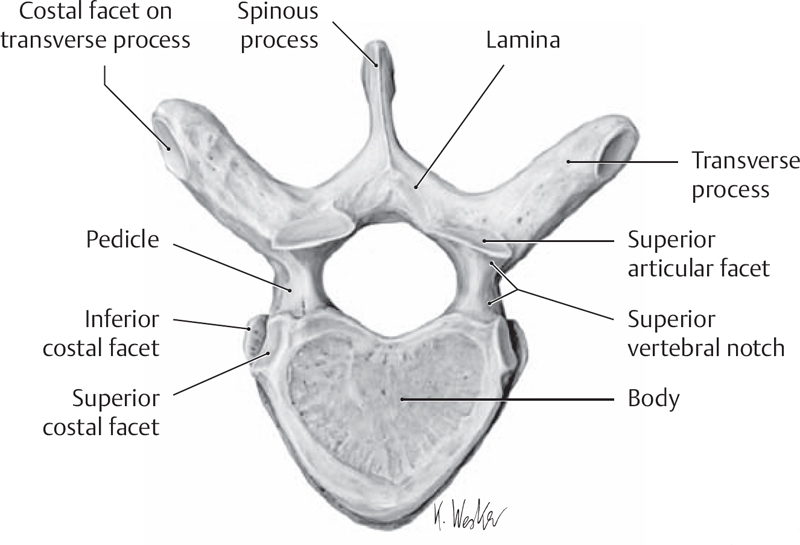
These bones help protect your spinal cord from injury while allowing you to twist and turn. Web In the thoracic vertebrae the transverse processes articulate with the ribs. The top thoracic vertebra T1 connects with C7 in the cervical spine above while the bottom thoracic vertebra T12 connects with L1 in the lumbar spine below.
They are thicker and larger than the cervical vertebrae but smaller than the lumbar vertebral bones. The T8 and T9 vertebrae are found at the same level as the xiphoid process. Seventh thoracic vertebrae T7.
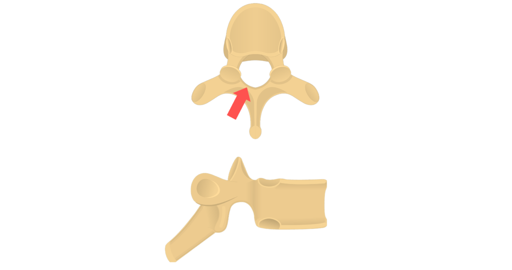
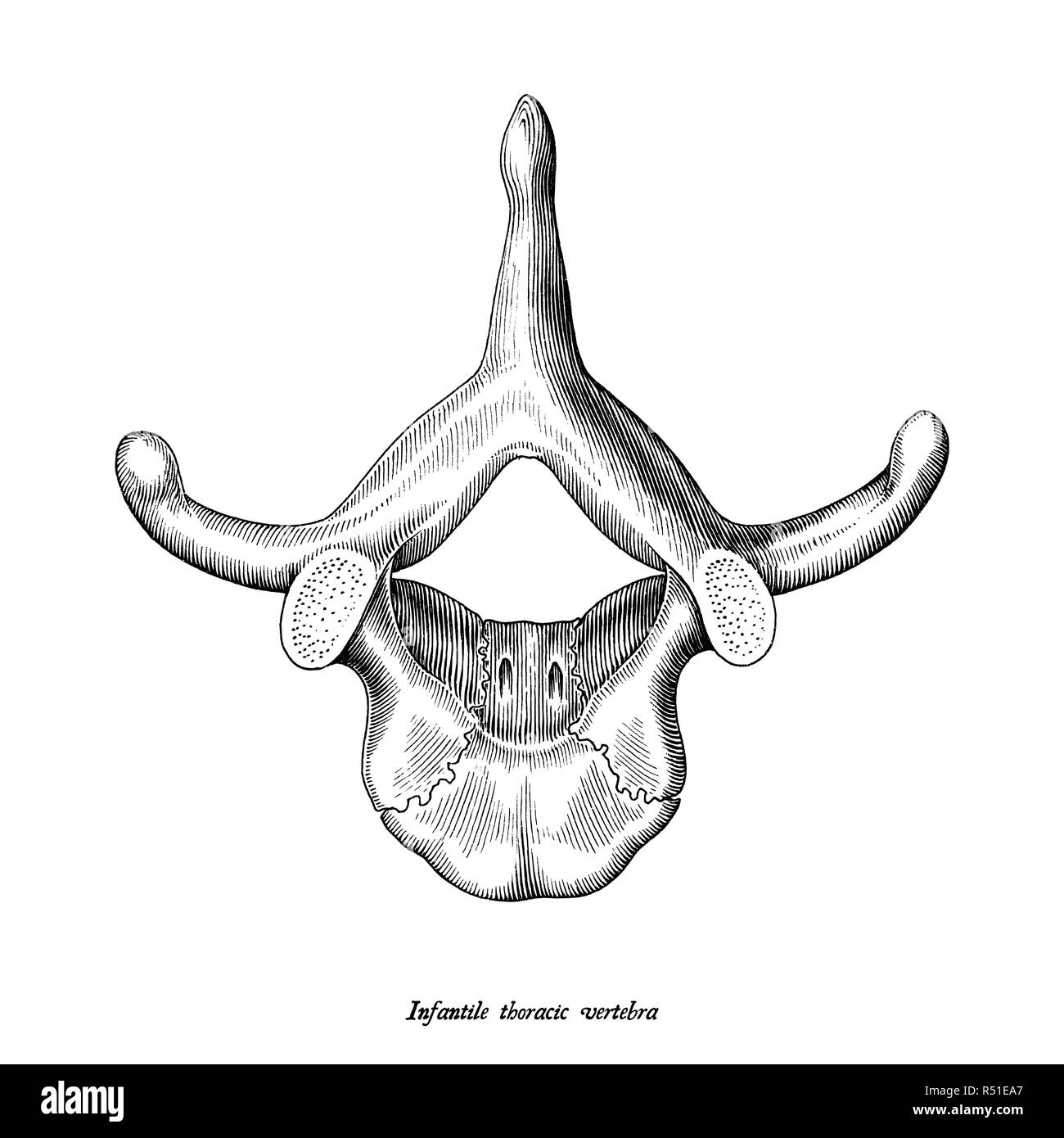
In addition to being connected to adjacent vertebrae the thoracic vertebrae are also connected to ribs. Thoracic vertebrae are unique among the bones of the spine in that they are the only vertebrae that support ribs and have overlapping spinous processes. It consists of 12 vertebrae that are distinct in.
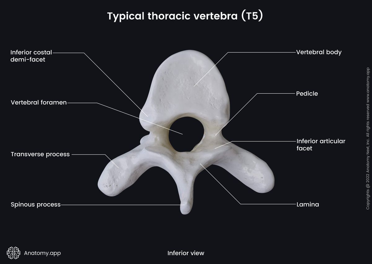
14mm shortest pedicle T10. The thoracic region contains 12 vertebrae denoted T1-T12. Lamina connect the transverse and spinous processes.
Web The thoracic cage rib cage forms the thorax chest portion of the body. The lumbar vertebrae are the largest movable bones of the. Along with the sternum and ribs the thoracic spine forms part of the thoracic cage.
Web Arachnoid Mater C1 Atlas - 1st Cervical Vertebra C2 Axis - 2nd Cervical Vertebra C3 3rd Cervical Vertebra C4 4th Cervical Vertebra C5 5th Cervical Vertebra C6 6th Cervical Vertebra C7 7th Cervical Vertebra Coccyx Dorsal Root of Spinal Nerve Dura Mater Fat in Epidural Space Iliolumbar Ligament Inferior Articular Process. Its part of the complex network of bones nerves and muscles that allow for the flexibility and movement of the upper back and neck. The characteristic feature for a typical midthoracic vertebra is the spinous process which is long and has a pronounced downward angle that causes it to overlap the next inferior vertebra.
Web The thoracic vertebra articulating with a pair of ribs. Eighth thoracic vertebrae T8. The thoracic cage protects the heart and lungs.
The first four T1-T4 and last four T9-T12 thoracic vertebrae share some characteristics with the. The ribs are anchored posteriorly to the 12 thoracic vertebrae T1T12. Pedicles connect the vertebral body to the transverse processes.
2 on the transverse processes and 4 demifacets. Web The T6 vertebra is the sixth bone in the thoracic spine located beneath the T5 vertebra. The thoracic spine sits between the.
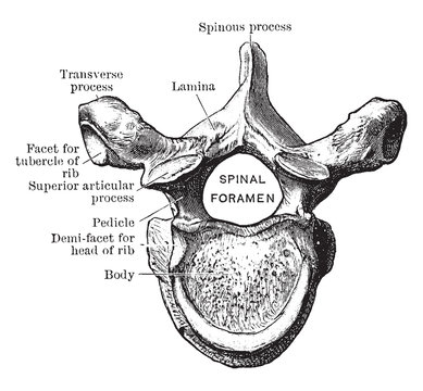
The facets of the transverse processes articulate with the tubercle of the associated rib. Web The thoracic spine is comprised of 12 vertebrae labeled T1 through T12. Web The sixth thoracic vertebrae T6 located just below the level of the shoulder blades works in conjunction with the remaining 11 segments to protect the nerves of the spine.
Seventh thoracic vertebra T7 The thoracic spinal nerve 7 T7 passes out underneath it. Web The seventh thoracic vertebra T7 is located in the mid to lower dorsal area at the inferior angle of the scapula shoulder blade. 15-17deg cephalad for majority of thoracic spine.
Varies from 10deg mid thoracic spine to 30deg L5 sagittal pedicle angle. All the thoracic vertebrae except the two at the bottom T11 and T12 form these articulations. Articulating with the ribs.
Learn vertebrae anatomy quicker and more efficiently with these diagrams and interactive quizzes. Web Sixth thoracic vertebrae T6. Web The thoracic spine has 12 vertebrae stacked on top of each other labeled from T1 down to T12.
Web The thoracic spine is part of the vertebral column that supports the chest area and provides posterior attachment for the ribs some thoracic wall muscles muscles of the upper limb abdomen and back. The bodies of the thoracic vertebrae are larger than those of cervical vertebrae Figure 726. The thoracic vertebrae are a group of twelve small bones that form the vertebral spine in the upper trunk.
Web Upper Back Upper Back The spine in the upper back and abdomen is known as the thoracic spine. Web Thoracic vertebrae contain several distinctive features. Web Your thoracic spine consists of 12 vertebrae labeled T1 through T12.
Articular processes form joints between one vertebra and its superior and inferior counterparts. Web T h o r a c i c S p i n e Functions Protecting the spinal cord and providing its passage through the tunnel at the center of the vertebral column as the vertebrae stack one after another. It is one of the three major sections of the spinal column.
Web The primary characteristic of the thoracic vertebrae is the presence of costal facets. See Vertebrae in the Vertebral Column. Costal facets that articulate with the ribs heart shaped vertebral bodies smaller vertebral foramina and long and strong spinous and transverse processes which point inferiorly.
It consists of twelve vertebrae which are separated by intervertebral discs. Eighth thoracic vertebra T8 The eighth thoracic vertebra is together with the ninth thoracic vertebra at the same level as the xiphisternum. Vertebrae are the 33 individual interlocking bones that form your spinal column.
The posterior superior and lateral views of a thoracic vertebra. Web The thoracic spinal nerve 6 T6 passes out underneath it. Web The thoracic vertebrae are bones located between the cervical and lumbar vertebrae.
Web The intervertebral discs are responsible for this mobility without sacrificing the supportive strength of the vertebral column. The intervertebral discs along with the laminae pedicles and articular processes of adjacent vertebrae create a space through which spinal nerves exit. Web The thoracic spine i s the second segment of the vertebral column located between the cervical and lumbar vertebral segments.
There are 6 facets per thoracic vertebrae. There are 12 thoracic vertebrae in humans and these bones increase in size as you move down the body.
Thoracic Vertebrae Anatomy And Labeled Diagram Getbodysmart
Anatomy Illustration Of The Thoracic Vertebrae Hi Res Stock Photography And Images Alamy
/images/vimeo_thumbnails/258798678/El29qHkkEoWw88WXJx8w_overlay.jpg)
Thoracic Vertebrae Anatomy Function And Definition Kenhub

Sixth Thoracic Vertebra From Above Vintage Vector Image
Thoracic Vertebrae Images Browse 12 965 Stock Photos Vectors And Video Adobe Stock

Typical Thoracic Vertebrae Radiology Reference Article Radiopaedia Org
![]()
Thoracic Vertebrae Anatomy Function And Definition Kenhub

Thoracic Vertebrae Wikipedia

Typical Thoracic Vertebra Diagram Medimartt Youtube

Vertebral Anatomy In The Florida Manatee Trichechus Manatus Latirostris A Developmental And Evolutionary Analysis Buchholtz 2007 The Anatomical Record Wiley Online Library

Pediagenosis Thoracic Vertebrae Thoracic Anatomy

Anatomy In Motion A Patient S Guide To Anatomy And Function Of The Spine By The University Of Maryland Medical Center Introduction The Spine Is One Of The Most Important Parts Of
Thoracic Vertebrae Encyclopedia Anatomy App Learn Anatomy 3d Models Articles And Quizzes
![]()
Thoracic Vertebrae Anatomy Function And Definition Kenhub

Thoracic Vertebrae An Overview Sciencedirect Topics
Thoracic Vertebrae Wikipedia
Thoracic Spine Neupsy Key